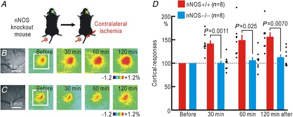
Figure 2. Cortical potentiation during hindpaw ischaemia contralateral to the stimulated hindpaw in wild‐type and nNOS knockout mice.

A, experimental method. B, cortical responses elicited by hindpaw stimulation in wild‐type (nNOS+/+) mice before and after the onset of hindpaw ischaemia. Original and pseudocolour images in ΔF/F 0 of the right somatosensory cortices are shown. C, cortical somatosensory responses elicited by hindpaw stimulation in knockout (nNOS−/−) mice before and after the onset of ischaemia in the right hindpaw. Original and pseudocolour images in ΔF/F 0 are shown. D, relative amplitudes of the cortical responses normalized by those recorded before the onset of hindpaw ischaemia. [Color figure can be viewed at http://wileyonlinelibrary.com]
