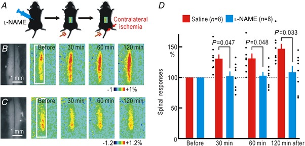
Figure 4. Spinal spreading potentiation during right hindpaw ischaemia with or without spinal application of l‐NAME.

A, experimental method. B, spinal responses in the left spinal cord elicited by brush vibration applied to the sole of the left hindpaw. In these mice, 5 μl saline was applied to the spinal cord before the recordings. Original and pseudocolour images in ΔF/F 0 of the spinal cord are shown. The time after the onset of ischaemia in the right hindpaw is shown in each pseudocolour image. The white rectangle in the left‐most pseudocolour image represents the ROI for the measurement of the response amplitude in ΔF/F 0. C, spinal responses elicited by left hindpaw stimulation before and after the onset of ischaemia in the right hindpaw. In these mice, l‐NAME (5 μl, 100 mM) was applied to the spinal cord. Original and pseudocolour images in ΔF/F 0 are shown. D, relative amplitudes of the spinal responses normalized by those recorded before the onset of hindpaw ischaemia. [Color figure can be viewed at http://wileyonlinelibrary.com]
