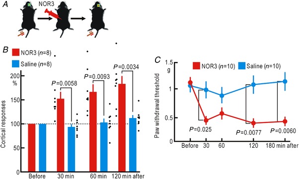
Figure 6. Cortical potentiation and mechanical hypersensitivity induced by spinal application of NOR3.

A, experimental method. B, relative amplitudes of the cortical responses normalized by those recorded before spinal application of 5 μl saline or 2 mM NOR3. The abscissa shows the time after the spinal application of saline or NOR3. C, mechanical threshold of the hindpaw withdrawal reflex measured by the von Frey test before spinal application of 5 μl saline or 2 mM NOR3. [Color figure can be viewed at http://wileyonlinelibrary.com]
