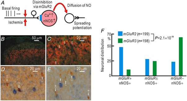
Figure 8. Colocalization of mGluR2 and nNOS in spinal neurons.

A, hypothetical relationship between signalling pathways mediated via mGluR2 and NO. B, immunohistochemical staining of group II mGluRs (mGluR2 and mGluR3, red) and nNOS (green). Colocalization of group II mGluRs and nNOS was observed in the superficial laminae of the dorsal horn. C, the same image shown in B at higher magnification. The cell bodies with nNOS (white arrows) were not stained with antibodies for group II mGluRs. D, immunostaining for nNOS (brown) and in situ hybridization of mRNA for mGluR2 (blue). Some cell bodies with nNOS were clearly (+), faintly (±) or not stained (–) with mRNA for mGluR2. White arrows show examples. E, immunostaining for nNOS (brown) and in situ hybridization of mRNA for mGluR3 (blue). F, relative distribution of nNOS‐positive spinal neurons that were clearly (+), faintly (±) or not stained (–) with mRNA for mGluR2 or mGluR3. [Color figure can be viewed at http://wileyonlinelibrary.com]
