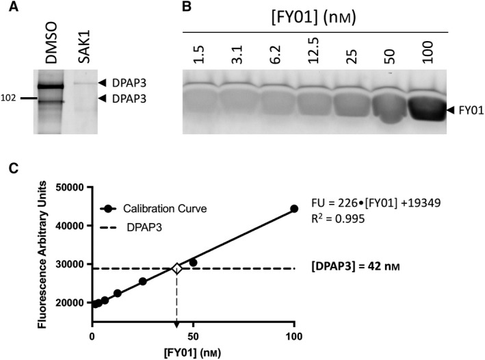
Figure 7.

Active site titration of DPAP3 using activity‐based probes. (A) Labelling of purified rDPAP3 by FY01 in the presence or absence of SAK1. Our stock of rDPAP3 was diluted 20‐fold in assay buffer, treated with DMSO or 1 μm SAK1 for 30 min, and labelled with FY01 for 1 h. Samples were run on an SDS/PAGE gel and DPAP3 labelling measured using a flatbed fluorescence scanner. (B) Calibration curve of free probe measured on the same SDS/PAGE gel as the one shown in A. (C) The fluorescence signal for rDPAP3 labelling and free probe was quantified using ImageJ, and the concentration of labelled rDPAP3 calculated based on the calibration curve.
