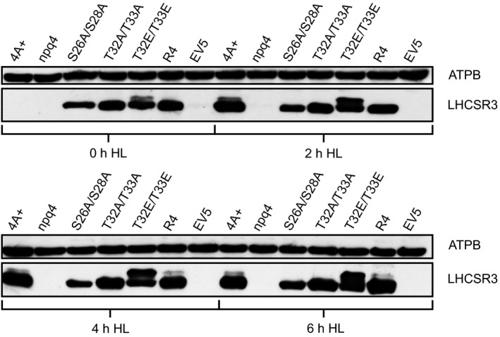
Figure 5.

Western blot analysis of whole cell extracts from cultures exposed to 0, 2, 4 or 6 h of 200 μmol photons m−2 sec−1 high light. Utilized strains were wild type 4A+, the LHCSR3 knockout strain (npq4), npq4 rescue strains with indicated amino acid alterations in the LHCSR3 protein, expressed under the control of the constitutive PSAD promotor, a rescue strain without amino acid alteration (R4) and an empty vector transformed control strain (EV5). Cultures were pre‐grown heterotrophically at ~20 μmol photons m−2 sec−1 low light and shifted to autotrophic high light conditions at the timepoint 0 h. For SDS‐PAGE, whole cell samples were adjusted to equal chlorophyll amounts. Immunodetection using an LHCSR3 specific antibody shows changes in abundance of the protein and varying migration behavior of different LHCSR3 species. Immunodetection of the β‐subunit of the chloroplast ATP synthase (ATPB) was used as a loading control.
