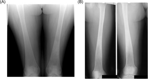
Figure 1.

Erlenmeyer flask deformity. (A) Typical appearance. Radiograph of the lower femora, showing the triangular outline of the metaphysis. Note the indistinct boundary between the cortex and medulla, typical of the Erlenmeyer flask deformity in Gaucher disease (GD). Incidentally, there is an area of serpiginous sclerosis in the left femoral metaphysis, suggestive of osteonecrosis. (B) Atypical appearance. Radiograph of the lower femora of a woman with GD who began enzyme replacement therapy at the age of 12 years. The proximal metaphysis has features similar to those in A, but the distal metaphysis has the more normal, trumpet‐shaped outline of the distal femur, with a clear border between the cortex and medulla. We speculate that the normal modeling process of endochondral ossification took place from the time of initiation of therapy
