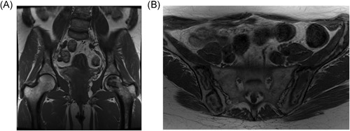
Figure 2.

Osteonecrosis. (A) Typical. T1‐weighted MR image of the pelvis, showing a geographic area of low signal in the head of the left femur. Note that the joint surface remains intact in this case; therefore, there is no deformity or degenerative change in the hip joint. (B) Atypical. T1‐weighted MR image of the pelvis in a different patient showing diffuse low geographic area of low and high signal through the pelvic bones bilaterally. The radiologic changes in (B) occurred gradually over years, on enzyme therapy, and without the typical symptoms of bone crisis
