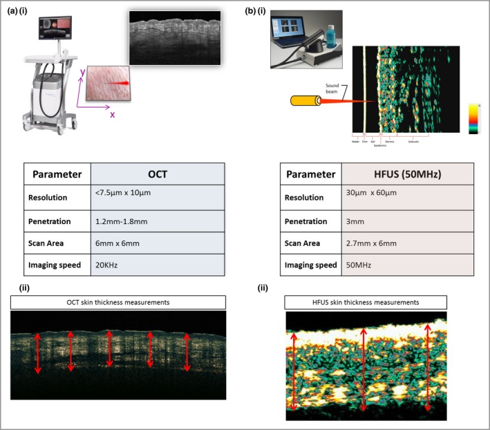
Figure 2.

Comparison of device parameters. (a) Optical coherence tomography (OCT) device. OCT uses low‐coherence near‐infrared radiation to produce an image of optical scattering from tissues, effectively creating an ‘optical ultrasound’ at a resolution close to that of histopathology. The device has a lateral optical resolution of < 7·5 μm and an axial resolution of 10 μm. The penetration depth is approximately 1·2–1·8 mm due to scattering effects, and the scan area is 6 × 6 mm. (b) High‐frequency ultrasound (HFUS) device. HFUS uses sound waves, and the ultrasonic waves are partially reflected at the boundary between adjacent structures, producing echoes of different amplitudes. The epidermis, dermis and subcutis are displayed in the image. We chose to use a 50‐MHz probe, which has a lower resolution of 30 × 60 μm than OCT and a greater penetration depth of 3 mm with a scan area of 2·7 × 6 mm.
