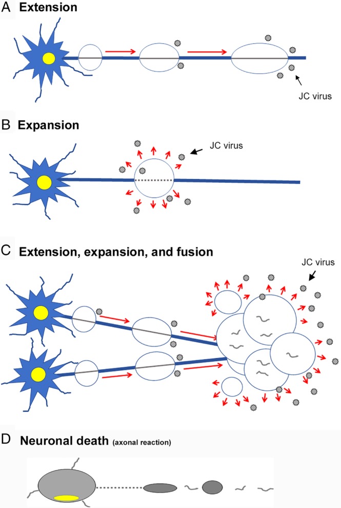
Figure 9.

Schematic diagrams showing pathologic processes of PML. How do the PML lesions spread in a tract‐dependent manner? JCV progenies likely spread along nerve axons. (A) In the extension stage, JCV forms proliferation foci along axons in a skipping manner. JCV‐infected cells are usually seen in the borders of extending lesions. (B) In the expansion stage, JCV proliferates locally, leading to expansion of the demyelinating areas. JCV‐infected cells are seen in the areas around expanding lesions. (C) In the fusion stage, the demyelinating lesions fuse with one another to form larger lesions. The brain tissue is severely damaged with axonal destruction. (D) In neuronal death stage, neurons die due to axonal destruction.
