Figure 4.

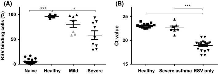
Eosinophils from asthma patients have a reduced capacity to bind and inactivate virus, but still inactivate virus in vivo (A). DiD‐labeled RSV determined by flow cytometry. Eosinophils without exposure to DiD‐labeled RSV (Naive), and after exposure to DiD‐labeled virus for eosinophils from 4 healthy (Healthy), 8 mild to moderate asthmatics (Mild asthma) and 9 severe asthmatics (Severe asthma) for 16 h. Eosinophils from mild asthma patients not on ICS are depicted in gray. B, Infectious RSV (depicted by Ct‐values) derived from lysates from RSV co‐incubated with eosinophils, reflected by 24‐h replication on epithelial cells. RSV only is RSV added directly to the epithelial cells (n = 16). Healthy refers to lysates from eosinophils from healthy donors (n = 4) and Severe asthma to those from severe asthma patients (n = 7). Ct value of 27.89 (5000 c/PCR) refers to a virus concentration of 6250 c/mL. Data are expressed as mean ± SEM. Paired or unpaired t test *P < 0.02, ***P ≤ 0.0001
