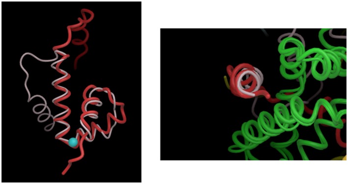
Figure 4.

Structural similarity between the Rb. sphaeroides ChrR‐ & E. coli RseA‐ASD. The left panel shows the structural similarity between helices I‐III and the displacement of helix IV of the ASD of Rb. sphaeroides ChrR (red) and E. coli RseA (white). The blue sphere is the Zn2+ atom in the ChrR‐ASD. The right panel shows that, despite this displacement of helix IV in the ASD of ChrR (red) and RseA (white), it interacts with a structurally conserved part of region 2 in the cognate Group IV sigma factors (region 2 of the Rb. sphaeroides and E. coli σE proteins are both shown in green). Figures modified from (Campbell et al., 2007).
