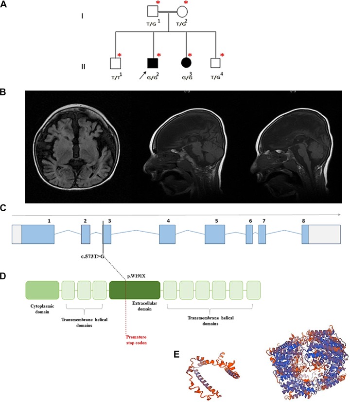
Figure 1.
A, The family pedigree of the affected siblings. B, Magnetic resonance imaging (MRI) findings for the affected male, at 4 years of age. Note the absent corpus callosum, the hypomyelination of the cerebral hemispheres and brain atrophy. C, The SLC1A4 transcript demonstrating the mutation site. D, The protein topological structure and site of the premature stop codon. E, The predicted mutated protein 3D structure modeled by SWISSMODEL/ExPASy prediction tool to the left, compared to the modeled wild-type protein (right).

