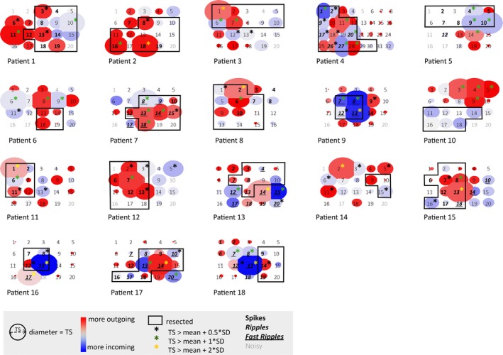
Figure 5.

Schematic representation of the total strength in the gamma band for all cases (1‐12 good outcome, 13‐18 poor outcome). The size of the circle represents the value of the total strength in the gamma band. The color indicates whether there are more/stronger outgoing (red) or incoming (blue) propagations. The resection (black rectangle) was based on results of preoperative examinations and tailoring based on spikes in the intraoperative electrocorticogram (ioECoG). Asterisks indicate nodes that should be included in the resection based on three different thresholds (black: total strength [TS] > mean + 0.5 SD, good outcome: 40 of 56 identified electrodes, poor outcome: 12 of 27 identified electrodes resected; green: TS > mean + 1 SD, good outcome: 26 of 35 identified electrodes, poor outcome: five of 14 identified electrodes resected; yellow: TS > mean + 2 SD, good outcome: five of five identified electrodes, poor outcome: three of seven identified electrodes resected; it should be noted that only five of 12 good outcome, and six of six poor outcome patients had electrodes with a gamma band total strength above mean + 2 SD). Different font styles indicate whether electrodes showed no events (regular), spikes (bold), ripples (bold italics), or fast ripples (bold italics underlined) in the epochs analyzed for network analyses or were removed because of noise (gray). What can be seen is that electrodes with a gamma band total strength above mean + SD threshold can be identified in all patients, and are most often included in the resected area in patients with good seizure outcome (1‐12) but not in patients with poor seizure outcome (13‐18). These are predominantly the channels that show events. The above method does not rely on the presence of epileptic activity in the epochs analyzed for network analysis. Patients 2, 11, and 12 did not have epileptic activity in their epochs, but did elsewhere in the ioECoG recording. Patient 3 had no events in the ioECoG overall, but a clear epileptic underlying substrate (focal cortical dysplasia 2A). However, the method does not yield significant results in tumor patients without any epileptic activity in their ioECoG recording overall (Patients 6, 10, and 14)
