Figure 2.

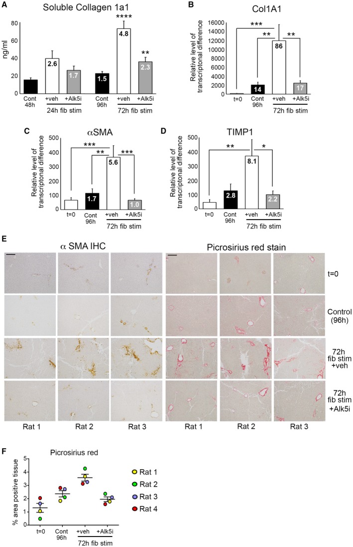
Modeling fibrosis and testing antifibrotic therapy in normal rat liver slices. (A) Graph shows soluble Col1a1 (ng/mL) levels in media of bioreactor‐cultured rPCLSs after 24‐hour rest and then 24‐ and 72‐hour culture ± fib stim (TGFβ1/PDGFββ) ± Alk5i (48‐ and 96‐hour total culture). (B‐D) mRNA levels of Col1A1, αSMA, and TIMP1 in rPCLSs at t = 0 and after 24‐hour rest + 72‐hour culture ± fib stim ± Alk5i (96‐hour total culture). (A‐D) Numbers on bars show fold change compared to t = 0. (E) Representative 100× magnification images of αSMA and picrosirius red–stained rPCLSs from three different livers at t = 0 and after 24‐hour rest + 72‐hour culture ± fib stim ± Alk5i. Scale bars = 200 µm. (F) Graph shows percentage area of picrosirius red–stained tissue in rPCLS at t = 0 and after 24‐hour rest + 72‐hour culture ± fib stim ± Alk5i. Data are mean ± SEM in n = 4 different livers. *P < 0.05; **P < 0.01; ***P < 0.001; ***P < 0.0001. Abbreviations: Cont, control; fib stim, fibrotic stimulation; IHC, immunohistochemistry; Veh, vehicle.
