
Figure 4.
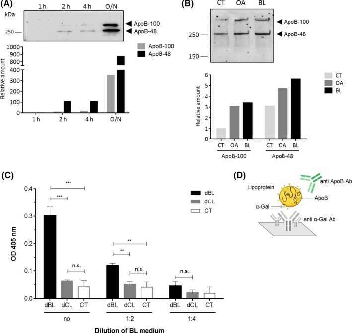
α‐Gal present in the basolateral medium is bound to chylomicrons. A, Anti‐ApoB immunoblot of basolateral media collected from Caco‐2 cells after 1 h, 2 h, 4 h, and overnight (O/N) incubation with undigested beef lipids. The graph below shows the relative amount of ApoB‐48 and ApoB‐100 at the different time points in comparison to the amount of the proteins produced after 1 h of incubation. B, Anti‐ApoB immunoblot of basolateral media collected from Caco‐2 cells incubated with only medium (CT), oleic acid (OA), and beef lipids (BL). Below, the relative amounts of ApoB‐48 and ApoB‐100 produced by the cells after being stimulated with OA and BL with respect to the amount produced by cells incubated with medium only (CT). C, Sandwich ELISA for detection of α‐Gal containing chylomicrons in the basolateral media of Caco‐2 cells that had been incubated with digested beef lipids (dBL), digested chicken lipids (dCL), or medium (CT). Media were applied either undiluted (no) or 1:2 or 1:4 diluted in PBS. In the sandwich ELISA, an anti‐α‐Gal antibody was used for catching and HRP‐labeled anti‐ApoB antibody for detection. Mean OD values indicating the binding of the anti‐ApoB antibody are shown on the y‐axis (**P ≤ 0.01, ***P ≤ 0.001, ns P > 0.05). D, Schematic representation of the ELISA set‐up