
Figure 2.
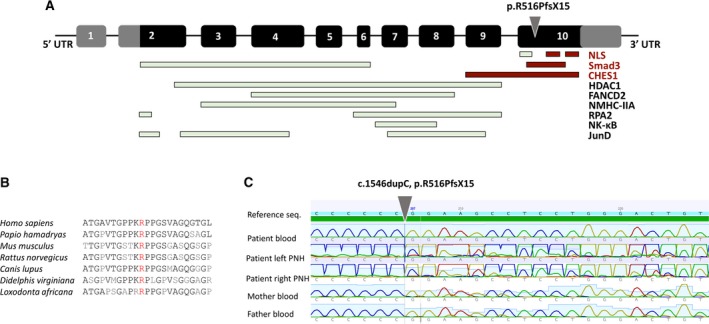
MEN1 gene schema and Sanger validation. A, MEN1 gene consists of 10 exons. Black panels represent transcribed and translated regions, whereas gray panels represent untranslated regions (UTRs). Exon 2 contains the start codon (ATG), and exon 10 the stop codon (TGA). Gray arrowhead indicates the location of the variant [c.1546dupC, p.R516PfsX15] found in the case presented in the article. NLS, nuclear localization signal. B, The affected variant is highly conserved, and the gene itself is highly conserved. Mouse Men1 demonstrates 97% identity/98% similarity to human MEN1 at the amino acid level.18 C, Sanger validation confirmed the absence of the variant in the peripheral blood DNA and the presence of the variant in a mosaic form in periventricular nodular heterotopia (PNH) on either side