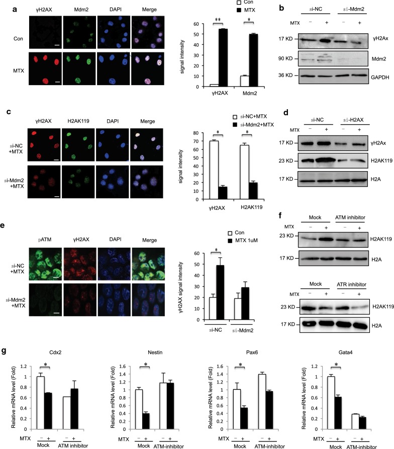
Fig. 4.
a Immunostaining using MDM2 antibodies formed discrete foci in mESC cells 12 h after exposure to MTX (1 μM) and colocalized with γH2AX. The analysis was confined to the transfected cells. The error bars represent the SEM. b F9 cells exposure to MTX (1 μM) 12 h. γH2AX were detected in MDM2-depleted F9 cells at levels comparable to those observed in control siRNA-treated cells and analyzed by western blot. c Immunostaining using H2AK119ub1 antibodies formed discrete foci in mESC cells 12 h after exposure to MTX (1 μM) and colocalized with γH2AX. The analysis was confined to the transfected cells. Error bars represent the SEM. d F9 cells exposure to MTX (1 μM) 12 h. H2AK119ub1 were detected in γH2AX -depleted F9 cells at levels comparable to those observed in control siRNA-treated cells and analyzed by western blot. e Normal foci for phosphorylated ATM were formed 12 h after exposure in MTX (1 μM) F9 cells in which MDM2 was downregulated. The analysis was confined to the transfected cells. Error bars represent the SEM. f F9 cells treatment with MTX (1 μM) 12 h and/or ATM inhibitor KU55933 (3 mM) for 12 h/ATR inhibitor 24 h and analyzed by western blot indicated antibody. g F9 cells treatment with MTX (1 μM) 12 h and/or ATM inhibitor KU55933 (3 mM) for 12 h/and expression levels of NTC-related genes Cdx2, Nestin, Pax6 and Gata4 analyzed by RT-qPCR

