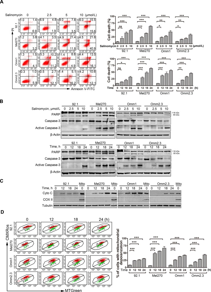
Fig. 2.
Salinomycin elicits apoptosis in UM cells. a Annexin V/PI apoptotic assay was performed in the UM cells treated with escalating concentrations of salinomycin for 24 h or at 10 μmol/L for various exposure time. Representative flow cytometry dot plots (left) for UM cells and quantitative analysis (right) from three independent experiments are shown. Data represent mean ± SD. ns, not significant; *, P < 0.05; **, P < 0.01; ***, P < 0.001, one-way ANOVA, post hoc comparisons, Tukey’s test. b Dose- and time-dependent of apoptosis-specific cleavage of PARP and caspase-3 activation was measured by Western blot after UM cells were incubated with increasing concentrations of salinomycin for 24 h or at 10 μmol/L for the indicated time periods. c UM cells were treated with 10 μmol/L salinomycin for different time, the levels of cytochrome c in the cytosolic extracts were analyzed by Western blot. COX II served as a mitochondrial content indicator. d UM cells were treated with 10 μmol/L salinomycin for the time indicated, and the mitochondrial potential was then detected by flow cytometry after dual staining with CMXRos and MTGreen. Data represent mean ± SD. ***, P < 0.001, one-way ANOVA, post hoc comparisons, Tukey’s test

