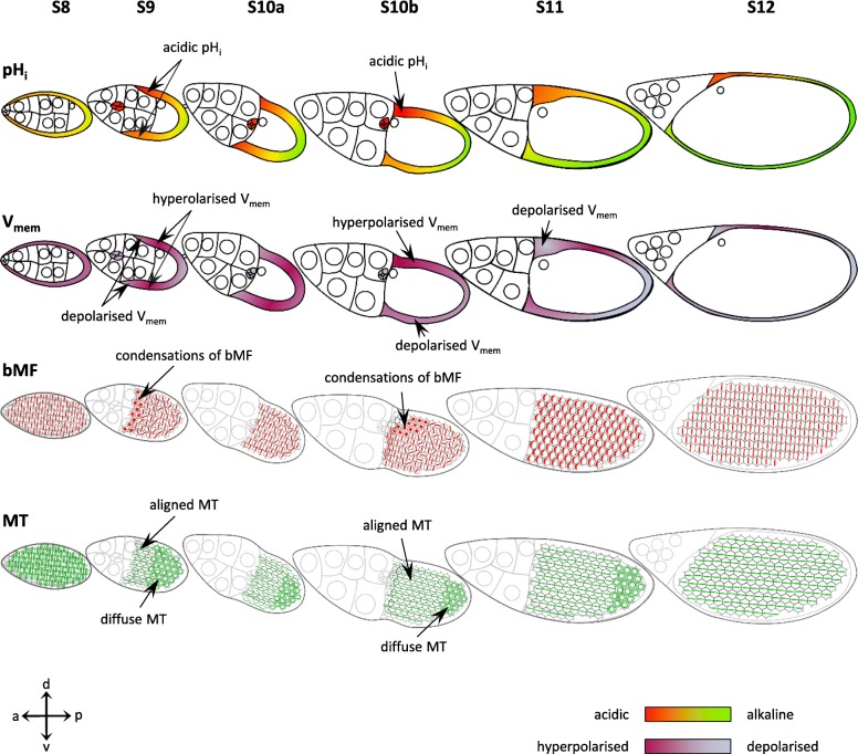
Fig. 6.
Changes in bioelectrical properties correlate with changes in cytoskeletal patterns in the FCE. Schematic drawings of follicles showing pHi and Vmem (according to [16]) and the cytoskeletal organisations (Figs. 4 and 5) in the FCE during S8–12. pHi: Beginning with S9, an a-p pHi-gradient develops with relatively acidic cFC and relatively alkaline pFC. From S10b onward, a d-v gradient establishes with relatively acidic dorsal FC and relatively alkaline ventral FC. Vmem: Beginning with S9, an a-p Vmem-gradient develops with relatively depolarised cFC and pFC, and relatively hyperpolarised mbFC. From S10b onward, a d-v gradient establishes with relatively hyperpolarised dorsal FC and relatively depolarised ventral FC. In late S10b/S11, the dorsal cFC and neighbouring FC become again more depolarised. bMF: In S8, the bMF in all FC are aligned in parallel perpendicular to the a-p axis. In S9, the bMF of flattening cFC condense and, in S10a, become aligned in parallel again. In dorsal cFC during S10b, condensation and subsequent disintegration of bMF occur, and this pattern spreads out toward pFC in S11. MT: The transversal orientation of MT in S8 changes during later stages: In S9, the MT of cFC become aligned along the a-p axis, whereas the MT of mbFC and pFC are diffusely organised. During S10a-12, the longitudinal orientation of MT spreads out toward pFC. The following correlations become obvious: FC showing condensed bMF (cFC in S9, dorsal cFC and neighbouring FC in late S10b/S11) are relatively acidic and relatively depolarised. Parallel alignment of bMF was observed in relatively alkaline FC, independent of Vmem (all FC in S8, mbFC and pFC in S9 and S10a, ventral mbFC and pFC in S10b, all FC in S12). Longitudinal orientation of MT was detected in more acidic FC, independent of Vmem (cFC in S9–12, dorsal mbFC in S10a-12), or in more alkaline FC with depolarised Vmem (ventral mbFC in S10a-12, pFC in S12)

