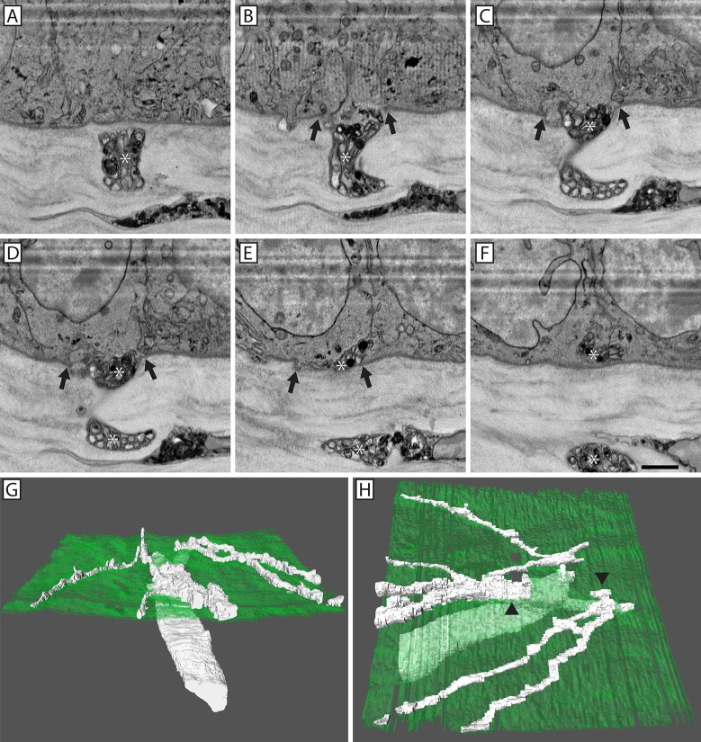
Fig 5. 3D reconstruction of nerve penetration through the epithelial basal lamina.
A series of SBF-SEM images showing a penetrating electron dense corneal nerve (*; A-F) that entered the epithelium through a discontinuity in the basal lamina (Panels B-E; arrows). A continuous basal lamina was present on either side of the penetration point (A & F). 3D reconstruction of the penetrating nerve (white) as it passed through the basal lamina (green; G & H). The nerve bifurcated prior to penetration (H; arrowheads). After penetrating into the corneal epithelium, both nerve branches underwent ramification. Scale bar = 2 μm.

