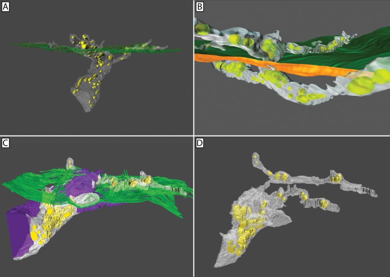
Fig 8. 3D reconstruction confirmed fusing axons lack mitochondria at the site of fusion.
Segmentation and 3D reconstruction of penetrating and fusing nerves (A-D). Mitochondria (yellow), penetrating axons (white), fusing axons (purple), and basal lamina (green) are shown. Conventional nerve penetration of the basal lamina involving multiple axons (A) or a single axon (B). In both cases, mitochondria were present throughout the nerve bundle on either side of the basal lamina. Mixed nerve bundle at the basal lamina showing penetrating and fusing axons (C). Mitochondria are clearly absent from the fusing axons. Isolation of the penetrating axons shows mitochondria to be distributed throughout the axoplasm (D).

