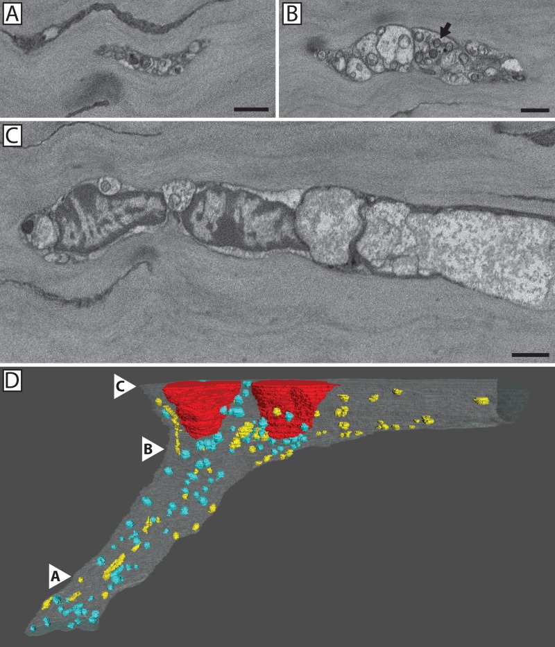
Fig 9. 3D reconstruction showed mitochondria are present within the distal portion of fusing axons.
Serial images show three levels (A-C) within the 3D reconstruction (D) of the distal portion of the mixed nerve bundle shown in Fig 4. The most distal portion of the nerve within the image series (A) was located ~60 μm distal to the site of fusion and it contained numerous mitochondria and an electron dense axoplasm. As the nerve bundle approached the fusion site, it increased in diameter (B & C). At ~35 um distance from the fusion site, mitochondria (blue) were no longer present in the fusing axons whereas mitochondria (yellow) were retained within the penetrating axons (D). White arrowheads denote the locations of panels A-C within the reconstructed nerve.

