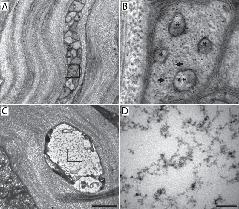
Fig 10. High resolution TEM showed an absence of microtubules in fusing neurons.
A conventional stromal nerve bundle (A) in which the inset is enlarged (B) to show cross-sectional views of microtubules identified by their size and distinctive hollow-ring appearance (arrows). Mitochondria are also present and identified by their double-membranes and internal cristae (*). A fusing nerve bundle (C) with an electron translucent axoplasm in which the inset is enlarged (D) to show the distinct lack of microtubules and mitochondria. Scale bar for panels A & C = 2 μm. Scale bar for panels B & D = 0.2 μm.

