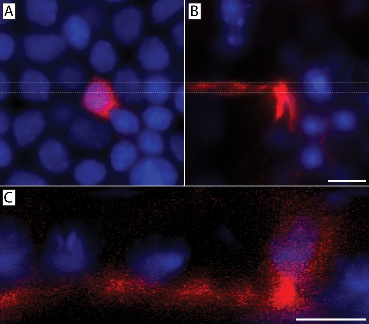
Fig 12. Orthogonal projection confirmed DiI transfer from corneal neuron to a single basal epithelial cell.
Two fluorescence images from a Z-stack showing a DiI (red) labeled basal epithelial cell (A) located above a DiI labeled stromal nerve (B). An orthogonal slice through the stack taken between the two dashed lines is shown in panel (C) where the DiI labeling extended uninterrupted from the neuronal plasma membrane into the epithelial cell membrane. DAPI (blue) staining denotes cell nuclei. Scale bars = 10 μm.

