
Figure 2.
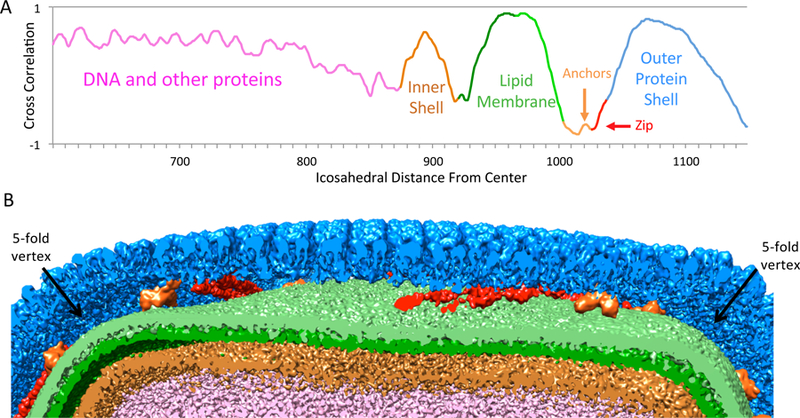
Layers in SGIV. (A) Radial plot (corrected for icosahedral & bulging geometry) showing peaks for inner shell, lipid membrane, anchor proteins, and outer protein shell. (See also Figure S1). (B) A cut-away view through the virion showing the different layers.