
Figure 3.
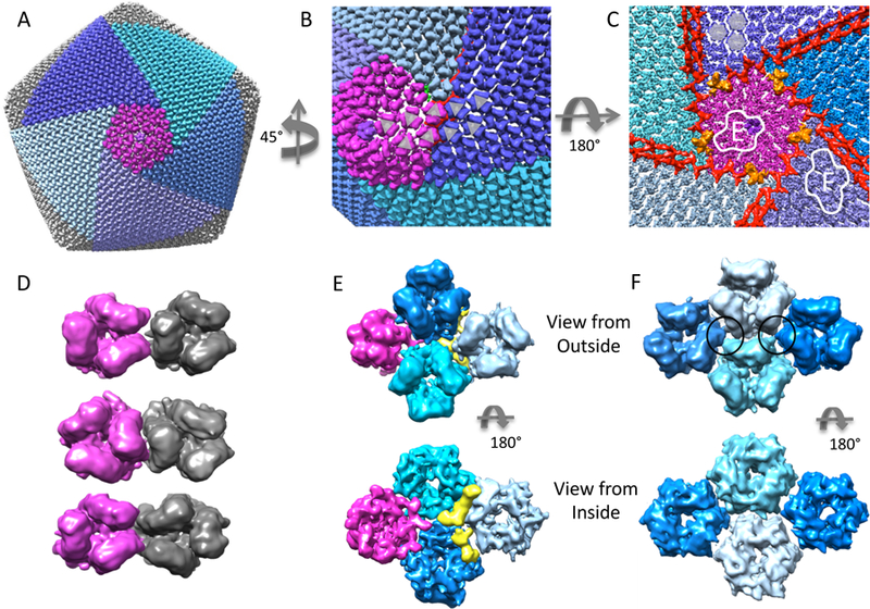
Outer coat proteins. (A) Surface of entire virion with a pentasymmetron (purple) and 5 trisymmetrons (blue/cyan) (see also Supplementary Movie 1). (B) Close up on the boundary between a pentasymmetron and trisymmetrons, showing a switch in orientation in the trimers that make up the coat. (C) View from inside, looking towards a pentasymmetron (purple). Anchor proteins are colored orange, and zip proteins are colored red. Two outlined sections are expanded in panels E and F. (D) Three different arrangements of neighbouring trimers: trisymmetrons only have the type shown at the top; within pentasymmetrons and penta-trisymmetron boundaries, all three types are seen. (E) Top and bottom views of 1 pentamer at the 5-fold vertex (purple) and 3 hexameric trimers (blue/cyan) taken from the pentasymmetron. The yellow segment may be a small protein or an unknown conformation for the N/C termini of the trimeric MCP. (F) Top and bottom view of 4 trimers taken from the center of a trisymmetron. Here, interactions between trimers (black circles) seem to occur mid-way between the inside and outside surfaces.