
Figure 5.
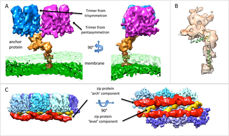
Anchor and Zip proteins. (A) Anchor protein (orange) shown next to bilayer lipid membrane, outer layer is transparent green, inner surface is solid green, and two of the capsomers it is adjacent to. The darker brown surface is the trans-membrane portion of the anchor protein. (B) Anchor protein, transparent surface, shown with the membrane protein P16 from PRD (PDB: 1W8X), showing similar architecture and trans-membrane helix. (C) Side and top view of zip proteins, red surfaces, connected to hexameric trimers, blue surfaces. The zip proteins appear to be composed of arch components, which arch away from the outer capsomeric surface, and level components, which are level with the inner surface of the capsomers.