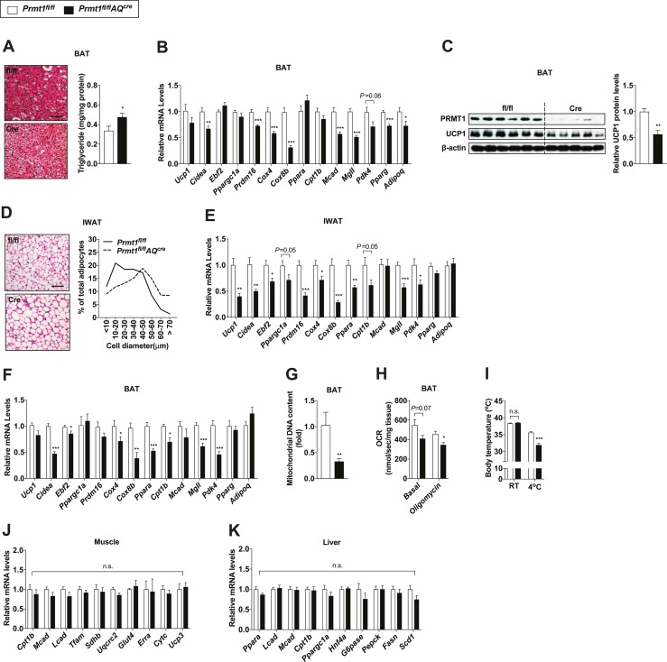
Figure 3.
Adipocyte-specific Prmt1-deficient mice display impaired adaptive thermogenesis. (A) H&E-stained images (left) and triglyceride levels of BAT (right; n = 8 for fl/fl, n = 12 for Cre) from Prmt1fl/fl and Prmt1fl/flAQcre mice exposed to cold (10°C) for 2 d. Scale bar, 50 μm. (B) qPCR analyses of thermogenic markers in BAT of Prmt1fl/fl (n = 8) and Prmt1fl/flAQcre (n = 7) mice after 2-d cold exposure (CE) (10°C). (C) Immunoblot analyses of PRMT1 and UCP1 (left) and quantification (right; n = 6 for fl/fl, n = 5 for Cre) of UCP1 levels in BAT of Prmt1fl/fl and Prmt1fl/flAQcre mice after 2-d CE (10°C). β-Actin was used as a loading control. (D) H&E-stained images (left) and distribution of adipocyte size (right) of IWAT from Prmt1fl/fl and Prmt1fl/flAQcre mice following 2-d CE (10°C). Scale bar, 50 μm. (E) qPCR analyses of thermogenic markers in IWAT of Prmt1fl/fl (n = 8) and Prmt1fl/flAQcre mice (n = 7) after 2-d CE (10°C). (F and G) qPCR analyses of (F) thermogenic gene expression (n = 6 for fl/fl, n = 7 for Cre) and (G) mitochondrial DNA copy number (n = 7 for fl/fl, n = 9 for Cre) in BAT from Prmt1fl/fl and Prmt1fl/flAQcre mice treated with 1 mg/kg/d CL 316,243 (CL) for 2 d at room temperature. Tissues were harvested 48 h after the first injection. (H) Oxygen consumption rate (OCR) in homogenates of BAT from Prmt1fl/fl and Prmt1fl/flAQcre mice pretreated with CL for 2 d and exposed to 4°C for 1 h (n = 6 per genotype). (I) Rectal body temperature of Prmt1fl/fl (n = 6) and Prmt1fl/flAQcre (n = 8) mice pretreated with CL for 2 d and exposed to 4°C for 1 h. (J and K) qPCR analyses of shivering- and lipid metabolism-related genes in (J) the skeletal muscle (n = 4 per genotype) and (K) liver (n = 9 for fl/fl, n = 7 for Cre), respectively, from Prmt1fl/fl and Prmt1fl/flAQcre mice exposed to cold (10°C) for 2 d. Data are presented as mean ± SEM. *P < 0.05; **P < 0.01; ***P < 0.005. n.s., not significant (P > 0.05).

