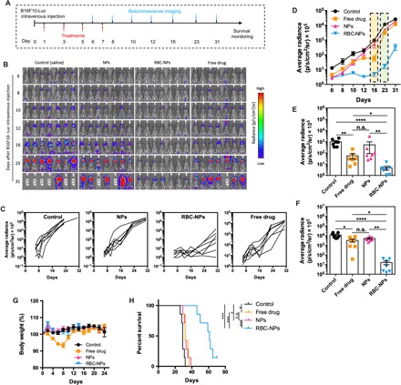
Fig. 4. The ELeCt platform inhibits lung metastasis progression and improves survival in the early-stage B16F10-Luc metastasis model.

(A) Schematic chart of the treatment schedule. (B) Bioluminescence images of lung metastasis at different time points. EXP indicates “Expired.” (C) Lung metastasis progression curve as depicted from in vivo bioluminescence signal intensity. (D) Quantification of lung metastasis burden at different time points (n = 7). (E) Scatter plot comparing the lung metastasis burden in different treatment groups as depicted from bioluminescence signal intensity on day 16 (n = 7). Significantly different (Kruskal-Wallis test): *P < 0.05, **P < 0.01, and ****P < 0.0001. (F) Scatter plot comparison of the lung metastasis burden on day 23 (n = 7). Significantly different (Kruskal-Wallis test): *P < 0.05, **P < 0.01, and ****P < 0.0001. (G) Body weight change of mice during the treatment period (n = 7). (H) Survival of mice under different treatments as displayed by Kaplan-Meier curves (n = 7). Significantly different (log-rank test): *P < 0.05 and ***P < 0.001.
