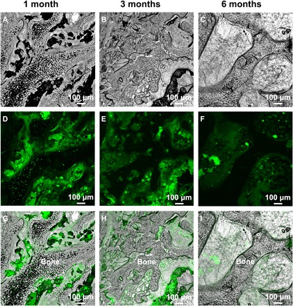
Fig. 3. LSCM images of bone tissue and HA-Tb particles.

LSCM images of bone tissue (A to C), HA-Tb particles with green fluorescence (D to F), and their overlapping images (G to I) after implantation for 1, 3, and 6 months. The images reveal the detailed interrelation between the HA-Tb material and bone tissue, and the distribution and amount change of the implanted HA-Tb material during bone reconstruction at the micro level.
