
Fig. 5. RvDp5 activates ALX/FPR2 to display dual anti-inflammatory and proresolving actions.
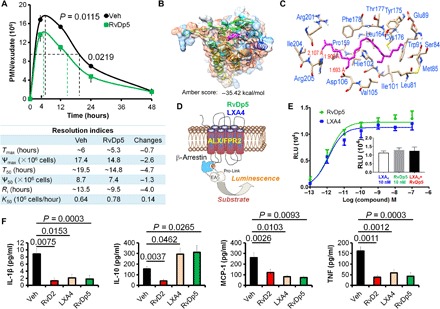
(A) Mice were injected intraperitoneally with Zym (1 mg) using PBS or RvDp5 (200 ng). Infiltrated PMNs were enumerated (upper panel), and resolution indices were calculated as indicated in Fig. 3B (bottom panel). (B) Three-dimensional (3D) chart of RvDp5 binding with ALX/FPR2. (C) Binding mode of RvDp5 structure ZINC35876755 in the binding pocket of ALX/FPR2. Important amino acid residues were shown, and the red lines indicated that the hydrogen bonds formed in the corresponding residues. (D) Diagrammatic principle of β-arrestin system. (E) Ligand (RvDp5 or LXA4)–receptor interaction was monitored in Chinese hamster ovary (CHO) cells using a β-arrestin system overexpressing ALX/FPR2. RLU, relative luminescence unit. (F) Mouse was treated intraperitoneally with Zym (1 mg/mouse; vehicle) with or without 200 ng of RvD2, LXA4, or RvDp5 for 4 hours, and the exudate levels of IL-1β, IL-10, MCP-1, and TNF were determined with ELISA. Error bars represent mean ± SEM. Two-tailed Student’s t test was applied for the P values.