Figure 4.

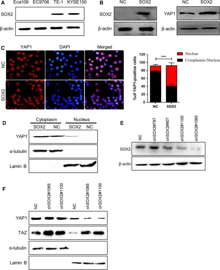
SOX2 controls endogenous YAP1 localization in ESCC cells. (A) Western blot analysis of endogenous SOX2 expression in a panel of ESCC cell lines. β‐Actin was served as a loading control. (B) Immunoblot analysis for SOX2 and total YAP1 protein levels after SOX2 overexpression in Eca109 cells. (C) Representative immunofluorescence staining and statistical analysis of YAP1 expression for the percentage of cells staining nuclear or both nuclear/cytoplasmic in Eca109‐vector and Eca109‐SOX2 cells. (D) Western blotting for nuclear YAP1 expression after SOX2 overexpression in Eca109 cells. α‐tubulin and Lamin B were used as loading controls. (E) Immunoblot analysis of SOX2 expression in KYSE150 cells after treatment with different shRNAs against SOX2. β‐actin was used as a loading control. (F) Western blotting for nuclear YAP1 and TAZ expression after SOX2 knockdown in KYSE150 cells. α‐tubulin and Lamin B were served as loading controls
