
Figure 2.
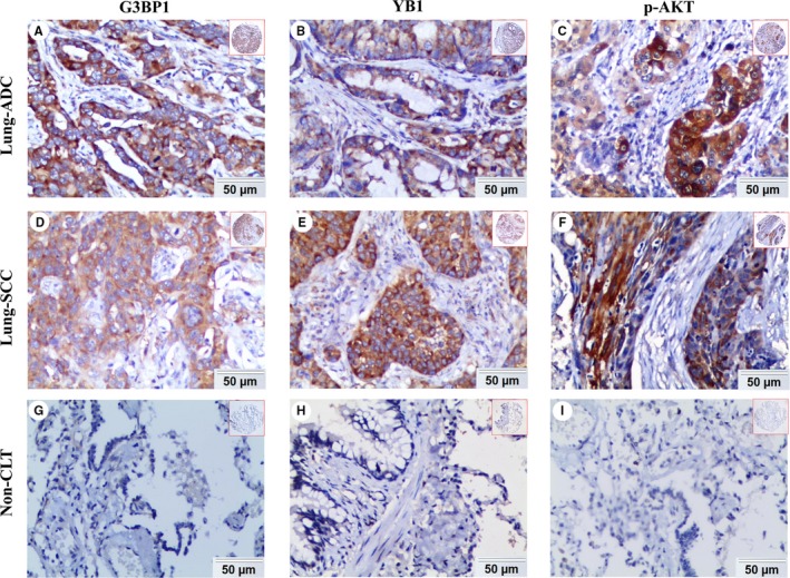
Expression of G3BP1, YB1, and p‐AKT proteins in lung ADC, lung SCC and Non‐CLT (noncancerous lung tissues) was detected by IHC. High expression of G3BP1 (A), YB1 (B), and p‐AKT (C) proteins was showed in lung ADC. High expression of G3BP1 (D), YB1 (E), and p‐AKT (F) proteins was also showed in lung SCC. Negative staining of G3BP1 (G), YB1 (H) and p‐AKT (I) proteins was found in noncancerous lung tissue. (IHC, DAB staining, original magnification 200× and 40×)