Figure 2.

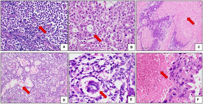
Histological features of EGGCTs. A, Seminoma. The neoplastic cells are arranged in solid lobules (red arrow) separated by thin fibrous septa containing lymphocytes. The cells are large‐sized, with abundant clear cytoplasm, roundish and relatively regular nucleus and prominent nucleolus. B, EC. The neoplastic cells are quite similar to seminoma cells, but the microscopic appearance is more variable. In this case, the architectural pattern is glandular (red arrow) and solid, and the neoplastic cells are more pleomorphic and atypical. C, EC. Coagulative necrosis (red arrow) is a diagnostic clue of EC. Attention must be paid to differentiating real coagulative necrosis from ischemic necrosis, a possible event in large or traumatized seminomas. D, YST. Relatively bland neoplastic cells arranged in the typical cystic architectural pattern (red arrow). E, YST. Schiller‐Duval bodies (red arrow) are a diagnostic clue, but they are present in about 50% of cases. F, Choriocarcinoma. The neoplastic cells are very large in size, with abundant slightly eosinophilic cytoplasm and atypical nuclei. Large hemorrhages are typically seen (red arrow). EC, embryonal carcinoma; EGGCTs, extragonadal germ cell tumors; YST, yolk sac tumor
