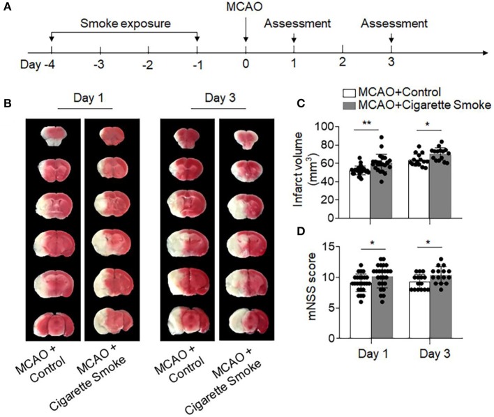
Figure 1.
Exposure to cigarette smoke-augmented neurodeficits and brain infarction after ischemia. (A) Schematic diagram illustrates experimental design. C57BL/6 mice were exposed to cigarette smoke for 4 consecutive days. Mice receiving normal air were used as controls. MCAO was performed at day 0. At days 1 and 3 after MCAO and reperfusion, neurodeficits and brain infarct volume were assessed. (B) TTC-stained brain sections from mice receiving cigarette smoke or normal air at indicated time points after MCAO and reperfusion. Summarized bar graphs show (C) infarct volume and (D) modified Neurological Severity Score (mNSS) in mice receiving cigarette smoke or normal air at indicated time points after MCAO and reperfusion. n = 25 per group at day 1, n = 15 per group at day 3. Mean ± SD. *P < 0.05, **P < 0.01.

