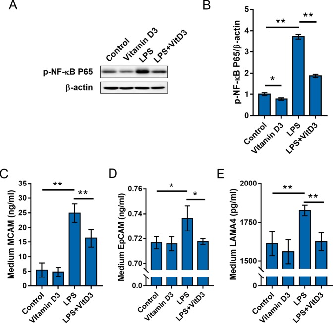
Figure 5.
The effects of vitamin D3 on LPS-stimulated NF-κB and adhesion molecules activation in RCC cells. (A,B) ACHN cells were pretreated with 1,25(OH)2D3, the active form of vitamin D3, and then stimulated with LPS for 6 h. Phosphorylated p65 was measured using Western blot. (A) A representative gel for p-p65 (upper panel) and β-actin (lower panel) was shown. (B) p-p65/β-actin. All experiments were repeated for six times. The grouping of blots cropped from different gels of same samples. All data were expressed as means ± S.E.M. (N = 6). **P < 0.01. (C–E) ACHN cells were pretreated with 1,25(OH)2D3, the active form of vitamin D3, and then stimulated with LPS for 6 h. The level of MCAM, LAMA4 and EpCAM in medium was measured using ELISA. (C) MCAM in medium; (D) LAMA4 in medium; (D) EpCAM in medium. All data were expressed as means ± S.E.M. (N = 8). **P < 0.01.

