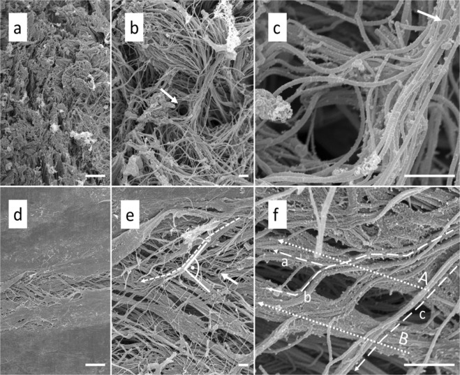
Figure 10.
Scanning electron microscopy of human temporal dura mater samples. (a) The arachnoid layer of the temporal dura mater reveals an isotropic organization at 1000x magnification (scale bar 10 μm). (b) 5000x magnification of a shows that the collagens run in multiple directions in the arachnoid layer (scale bar 1 μm). The arrow points to a channel that might form the bed for a vessel in the native state. (c) The channel depicted in b is shown at a higher magnification of 25,000 × (scale bar 1 μm). Note the bending of the collagen fibres (arrow) in proximity to the vessel channel. (d) The bone surface layer of the human temporal dura reveals a clear organization of the collagens at 1000x magnification (scale bar 10 μm). (e) A 5000x magnification of d displays the anisotropic structure of the bone surface layer (scale bar 1 μm). Collagen bundles are running in a preferred direction. The bone surface layer of the temporal dura is composed of several superimposed collagen layers which run into different directions with angles up to 90°. The arrow marks a single fiber, which is most likely elastin due to its single course crossing several subjacent collagen bundles (for the characteristics of elastic fibres in the SEM see Woplers47). (f) A 25,000x magnification of e shows the splitting of the collagen bundle marked with a dotted line in e (scale bar 1 μm). Note how the fibres of the splitting bundle either join collagen bundles A (dotted line a) and B (dotted line b) or just pass the bundles on their way (dotted line c).

