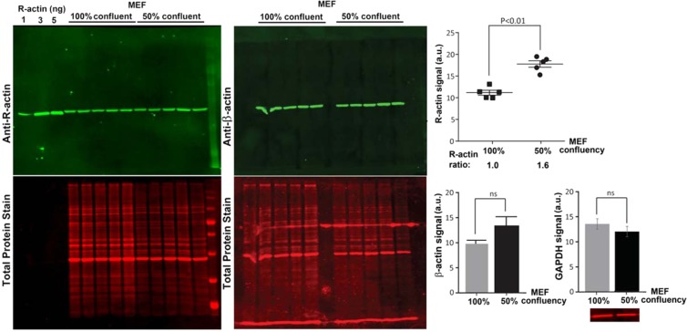
Figure 2.
Percentage of actin arginylation partially depends on cell confluency. Left, Western blots of MEF cells with different confluency probed with antibodies against R-actin and β-actin as marked. Right top, quantification of total fluorescence from the R-actin band, normalized according to signal of total protein staining. Right bottom, quantification of β-actin and GAPDH in the same samples. Inset on the bottom right shows the representative GAPDH signal used for quantification. Error bars represent SEM, n = 5 for actin and 3 for GAPDH, Student’s t-test was used to determine the P value.

