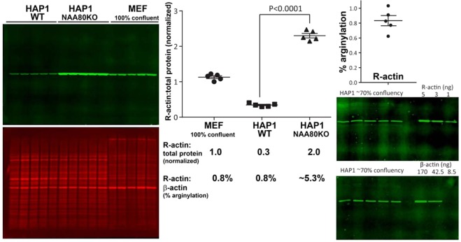
Figure 6.
Lack of N-terminal actin acetylation in HAP1 cells leads to a dramatic increase in N-terminal β-actin arginylation level. Left, Western blots of HAP1 WT, HAP1 NAA80 knockout and 100% confluent MEF cells. Middle, quantification and comparison of the R-actin fluorescence signal from these different cell types. Signal of total protein stain was used as reference for R-actin normalization. The 5.3% arginylation in HAP1NAA80KO cells is shown as an estimate, since no β-actin signal can be detected in these cells. Right, quantification of the percentage of R-actin in wild type HAP1. Error bars represent SEM, n = 5, Student’s t-test was used to determine the P value.

