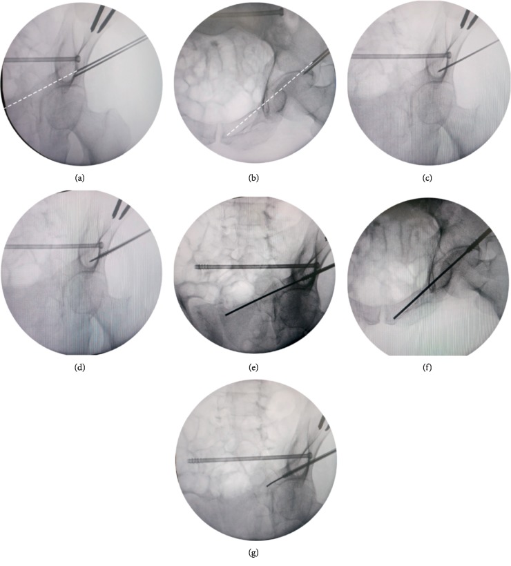
Figure 3.
Photographs demonstrating the insertion of antegrade superior ramus screws using the “blunt end” KW technique. (a-b) The position of the inferior KW inserted through the centered hole of the guide tunnel was not ideal, and then a second KW was inserted through 1 of the peripheral holes of the “plum blossom” until an ideal position was obtained (white dotted line). (c) Obturator outlet view was taken to confirm the position was good after inserting the KW 1-2 cm into the bone. (d) The preselected cannulated screw was inserted along the wire. (e-f) The “blunt end” KW technique was used again to complete the insertion of the guide wire under the surveillance of intraoperative fluoroscopic pelvic inlet and obturator outlet views. (g) The “sea snake head” technique was used so that the curved tip of KW could pass through the narrow corridor or broken end of the fracture smoothly.

