Fig. 2.

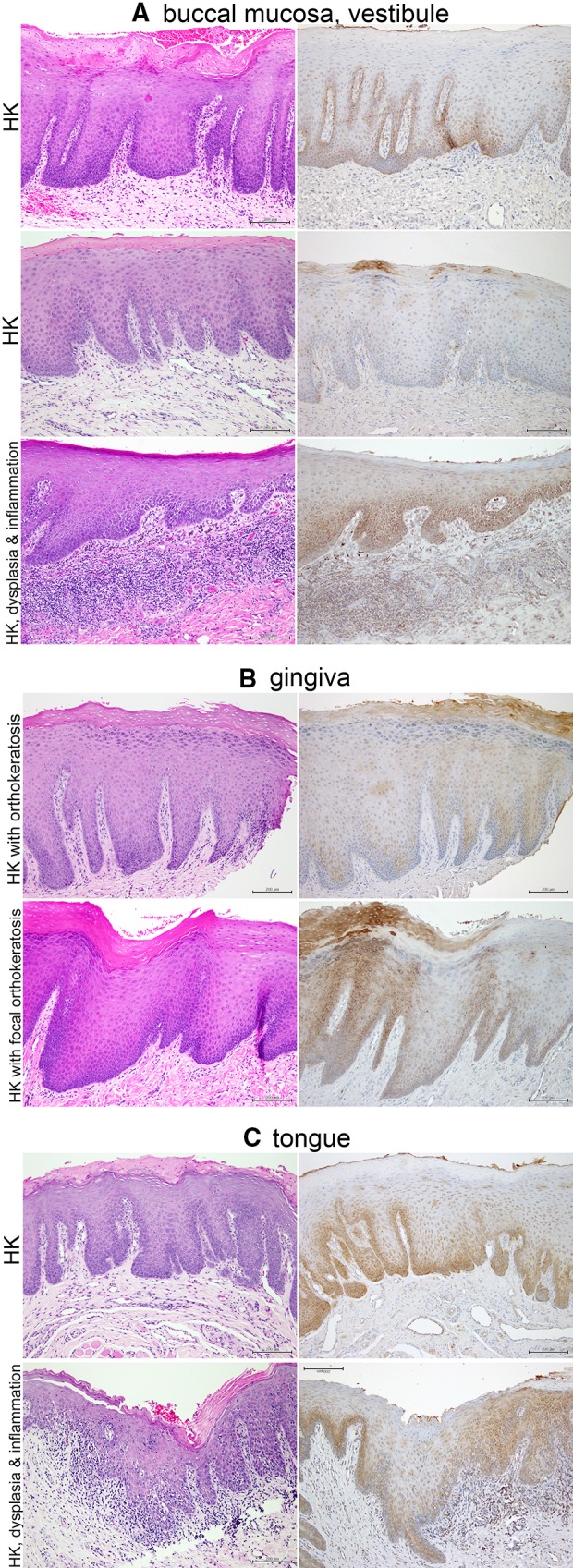
Representative examples of hyperkeratosis in the buccal mucosa or vestibule, gingiva and tongue (mag. ×100). a Buccal mucosa and vestibule. Left column—H&E, right column—TLR2. Note acanthosis and hyperparakeratosis (top two examples) and focal orthokeratosis (middle), with primarily basal and parabasal TLR2 expression. Small foci of perinuclear TLR2 expression are present in the keratinocytes of the spinous layer (middle). A case of hypeparakeratosis with focal acanthosis and with mild dysplasia (lower panel) shows retention of diffuse cytoplasmic TLR2 expression above the basal/parabasal layers, following the pattern of dysplasia. b Gingiva. Acanthosis, hyperpara- and orthokeratosis, and variable TLR2 expression. c Tongue. Acanthosis and hyperparakeratosis with TLR2 predominantly in basal and parabasal layers. Only small perinuclear foci of TLR2 appear to be present in the spinous layers. However, in the case of transition to moderate dysplasia (lower panel), note retention of diffuse cytoplasmic TLR2 expression in the spinous layers, following the dysplasia. Note: same portions of the specimens were photographed, although the H&E-stained and IHC-stained sections were not serial
