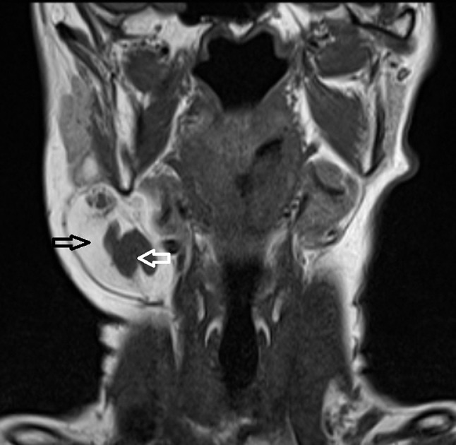
Fig. 1.

T1-weighted magnetic resonance imaging of head and neck. T1w coronal image showed an exophytic mass extending inferiorly from the tail of right parotid. The mass demonstrated T1w high signal (black open arrow), which was isointense to fat and there was an additional lobulated T1w isointense component (white open arrow)
