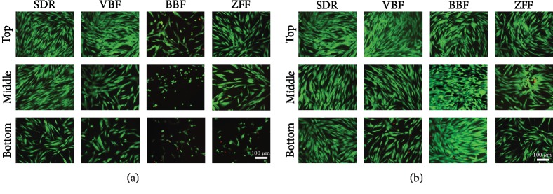
Figure 2.
Live and dead staining of human dental pulp cells (hDPSCs) incubated with (a) 100% or (b) 12.5% elute from different specimen depths for 24 h. Live/dead cells were stained green/red, respectively, and representative images were shown (n = 6). Bottom specimens showed fewer live cells in all groups for 100% elute. BBF and ZFF yielded more live cells than SDR and VBF at 100% elute. The number of live cells generally increased from 100% to 12.5% concentrations. Representative data are shown after triplicate experiments.

