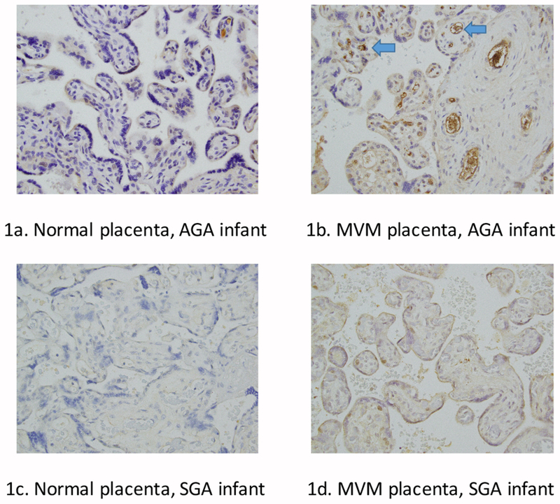
Figure 1.

normal placenta, AGA infant shows minimal AK staining. Figure 1b: MVM placenta, AGA infant shows increased endothelial AK staining (blue arrows), suggesting placental endothelial AK plays a protective role in intrauterine growth restriction. Figure 1c: Normal placenta, SGA infant shows low staining of AK in the endothelial cells. Figure 1d: MVM, SGA infant shows low endothelial staining of AK. In the MVM placental slides, distal villous hypoplasia, small terminal villi, and syncytial knots are also apparent.
