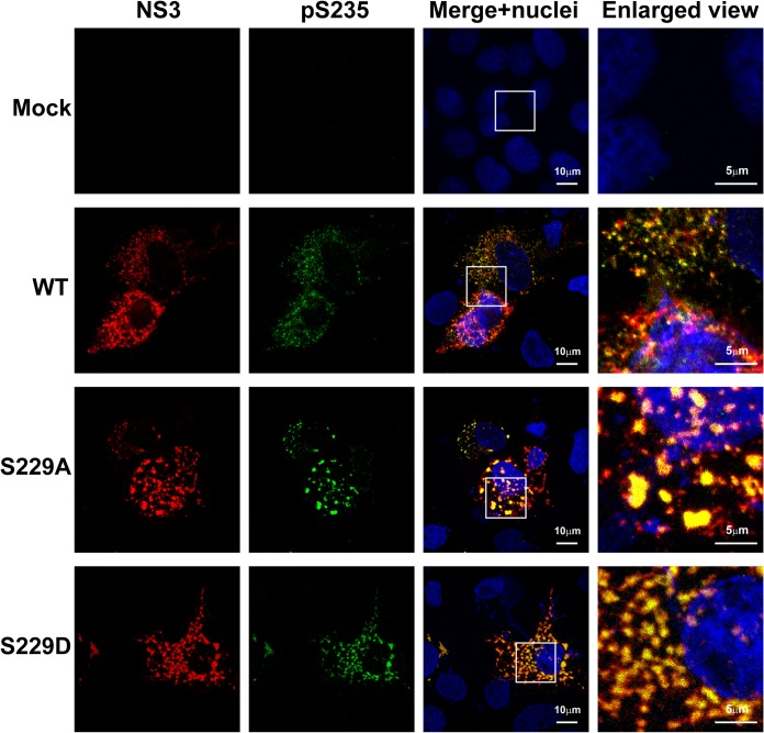
FIG 6.
S229A and S229D mutations altered intracellular distribution of NS5A with regard to NS3. Confocal immunofluorescence micrographs of NS3 (red) costained with S235 phosphorylated NS5A (pS235, green) in the T7-Huh7 cells transfected with wild-type (WT), S229A, or S229D mutant NS3-NS5B mutant constructs. The nuclei were stained with DAPI (blue). Boxes indicate areas enlarged.

