FIG 7.
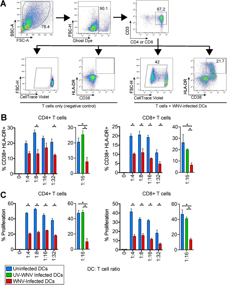
WNV-infected DCs are compromised in T cell proliferation. moDCs were left uninfected or infected with WNV (MOI of 10) for 24 h. Allogeneic CD4 or CD8 T cells were labeled with CellTrace violet (CTV) and incubated with uninfected or WNV-infected moDCs at the indicated DC/T cell ratios in the presence of an anti-E16 WNV blocking antibody to limit spreading of infection (5 μg/ml) for 6 days. (A) Representative flow cytometry gating strategy for proliferation by CellTrace violet as well as CD38 and HLA-DR positivity in CD4+ and CD8+ T cells. FSC-A, forward scatter area; FSC-H, forward scatter height; SSC-A, side scatter area. (B) The percentage of cells double positive for the T cell activation markers CD38 and HLA-DR on day 6 of allogeneic coculture of mock-, WNV-, or UV-WNV-infected moDCs with T cells. (C) The percentage of cells that had proliferated by day 6 of allogeneic coculture of mock-, WNV-, or UV-WNV-infected moDCs with T cells. The calculation of percent proliferation included any cell that diluted CTV compared to the level in a no-DC, T cell-only control. *, P < 0.05 (one-way and two-way ANOVA).

