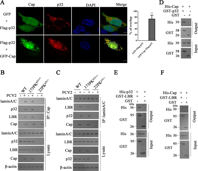
FIG 3.
p32 mediated the interaction of lamin A/C, lamin B receptor (LBR), and Cap. (A) PK-15 cells were transfected with pEGFP-Cap and pCI-p32-Flag vectors or with pCI-p32-Flag and pEGFP-N1 vectors; the cells were fixed and subjected to laser scanning confocal microscopy. Images represent the subcellular locations of green fluorescent protein (GFP)-Cap and Flag-p32 proteins (left), and histograms represent the percentage of overlap of Flag-p32 proteins with GFP-Cap, performed using ImageJ software and based on ≥15 cells/sample (right). **, P < 0.01 (compared with pCI-p32-Flag and pEGFP-N1 vector cotransfected cells). (B, C) p32 mediates the interaction of lamin A/C, LBR, and Cap protein. Wild-type PK-15, 173PKp32+/+, and 22PKp32−/− cells were infected with PCV2, and immunoprecipitation was performed to detect the Cap interaction with lamin A/C, LBR, and p32 using anti-Cap antibodies (B) or anti-lamin A/C antibodies (C). (D to F) Direct interaction of p32 with PCV2 Cap or LBR. Bacterially purified GST-p32 or glutathione S-transferase (GST) alone was incubated with purified His-Cap, and proteins bound to glutathione Sepharose beads were analyzed by immunoblotting with the indicated antibodies (D); purified GST-LBR or GST alone was incubated with purified His-p32 (E) or His-Cap (F). Proteins bound to glutathione Sepharose beads were analyzed by immunoblotting with the indicated antibodies.

