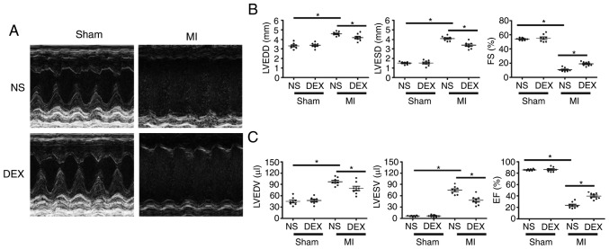
Figure 1.
Functional analysis of the left ventricle with echocardiography. (A) Representative M-mode echocardiograms from the Sham + NS, Sham + DEX, MI + NS, and MI + DEX mice. (B) The mean left ventricular LVEDD, LVESD and FS in the Sham + NS, Sham + DEX, MI + NS, and MI + DEX-treated mice. (C) The mean left ventricular LVEDV, LVESV and EF in the Sham + NS, Sham + DEX, MI + NS, and MI + DEX mice. Data are presented as mean ± standard error of the mean. n=6. *P<0.05. NS, normal saline; DEX, dexmedetomidine; MI, myocardial infarction; LVEDD, left ventricle end-diastolic dimension; LVESD, LV end-systolic dimension; FS, fractional shortening.

