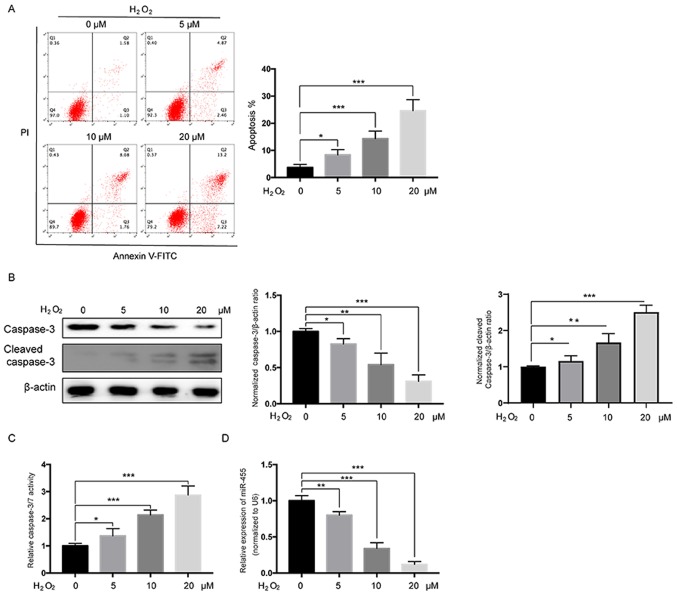
Figure 1.
H2O2 induces apoptosis and affects the expression of miR-455 in HESCs. (A) Flow cytometry analysis of the apoptosis rates in HESCs exposed to various doses of H2O2 for 24 h. (B) HESCs were treated with various concentrations of H2O2 for 24 h, and then, cellular lysates were collected and subjected to western blot analysis with the caspase-3 antibody. The histograms (right) show the densitometric analysis of the caspase-3 and cleaved caspase-3 western blot results. (C) HESCs were treated with various concentrations of H2O2, after which total cellular lysates were collected and subjected to a caspase-3/7 activity assay. (D) HESCs were treated with various concentrations of H2O2 for 24 h, and then, the levels of miR-455 were measured by qPCR. All data are shown as the mean ± SD of three independent experiments. *P<0.05, **P<0.01 and ***P<0.001. HESCs, human endometrial stromal cells; H2O2, hydrogen peroxide.

